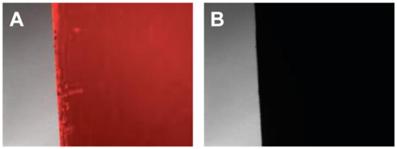
Figure 2.

Fluorescence microscopy images (40× magnification; 730 μm × 510 μm) of the edge of a stainless steel substrate coated with a polymer 1/DNA film 8-bilayers thick, incubated in an aqueous ethidium bromide solution that intercalates DNA and labels it with a red color (A) before electrochemically-induced erosion, and (B) after applying a potential of -1.1 V for 1 min in PBS.
