
Figure 1.
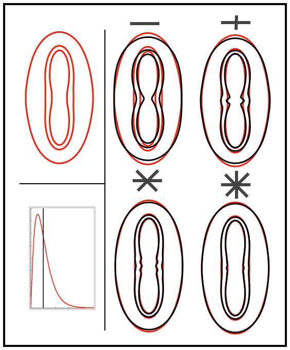
Illustration of the over-fitting using two-dimensional simulations. Top left: signal profile for a single fibre model using Gamma distributed diffusivities and 3 b-values = (1,2 and 3)×103 s/mm2. Right panel: black lines are maximum likelihood fits of the mono-exponential model with increasing number of crossing fibres (1,2,3 and 4). Notice how increasing the number of fibres artificially moulds the predictions to the simulated data, even though the data was generated using a single fibre. Bottom left: distribution of diffusivities for the red (Gamma) and black (dirac) signals.