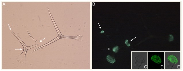
FIGURE 1.
Immunolocalization of mannans in isolated Arabidopsis trichomes. (A) Bright field and (B) epifluorescence micrographs of intact and broken trichomes isolated using the procedure of Marks et al. (2008). Arrows denote the locations of trichome branch breakpoints. (C–E) Confocal average projections of the base region of an isolated Arabidopsis trichome. For (B, D, E), the LM21 and LM22 antibodies (Marcus et al., 2010) were used for mannan immunolocalization.

