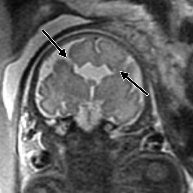
Figure 4a:

Bifrontal open-lip schizencephalies detected with fetal MR imaging at 33 gestational weeks. (a) Fetal coronal single-shot fast SE T2-weighted image demonstrates bilateral clefts (arrows) in the parenchyma that extend from the ventricular lumen to the subarachnoid space and are lined by signal intensity similar to that of the developing cortex. Findings are compatible with open-lip schizencephalic defects. Septal leaves are absent. (b) Corresponding coronal, three-dimensional, spoiled gradient-recalled acquisition in the steady state, T1-weighted image obtained at 1 year of age confirms the bilateral open-lip schizencephalies (arrows). (c) Fetal axial single-shot fast SE T2-weighted image also demonstrates the bilateral open-lip schizencephalic defects (arrows), as well as abnormal infoldings in the adjacent cortex. (d) Corresponding axial T1-weighted image obtained at 1 year of age confirms the bilateral open-lip schizencephalies (arrows), as well as adjacent polymicrogyria.
