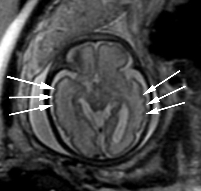
Figure 5c:

Polymicrogyria detected with fetal MR imaging at 30 gestational weeks. (a) Coronal single-shot fast SE T2-weighted image in a 25-week-old fetus demonstrates normal sulcation pattern without any evidence of developing polymicrogyria. (b) Coronal single-shot fast SE T2-weighted image in the same fetus at 30 gestational weeks demonstrates shallow appearance of the frontal sulci (arrows). (c) Axial single-shot fast SE T2-weighted image at 30 gestational weeks also demonstrates abnormal infoldings of the temporal cortex bilaterally, consistent with polymicrogyria (arrows). (d) Postnatal coronal three-dimensional fast SE T2-weighted image obtained at 3 months of age confirms the findings from 30-week fetal MR image with bifrontal polymicrogyria. Abnormal T2 hyperintensity in the white matter is also noted (and was not detected with fetal MR imaging). (e) Postnatal axial SE T2-weighted image also demonstrates bilateral temporal lobe polymicrogyria, as detected with fetal MR imaging.
