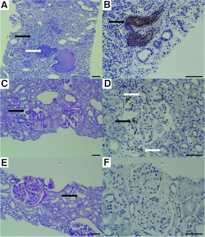
Figure 4.
Histologic findings in periodic acid Schiff reaction (PAS) staining (×100; scale bars, 100 μm) and immunohistochemistry with an antibody to the S100A8/A9 heterocomplex (×200, brown; scale bars, 100 μm) in pyelonephritis (A and B; urinary calprotectin, 8232 ng/ml), lupus nephritis (C and D; urinary calprotectin, 1422 ng/ml), and a patient fulfilling the clinical criteria of prerenal disease (E and F; urinary calprotectin 3 ng/ml). (A) PAS staining in pyelonephritis reveals marked tubulointerstitial nephritis, patchy interstitial inflammation by polymorphonuclear leukocytes (black arrow), and acute tubular injury with a dense inflammatory infiltrate (white arrow). (B) Neutrophils occurring in tubular microabscesses are strongly positive in the calprotectin staining (black arrow). (C) PAS staining in lupus nephritis shows two glomeruli with mild to moderate mesangial hypercellularity segmentally involving the glomerular tuft and increasing the mesangial matrix. Moreover, there is a small segmental lesion with endocapillary proliferation (black arrow). Tubular injury is mild with focal atrophy of tubular epithelial cells. (D) Calprotectin staining evoked positive signals in both the intraglomerular space (black arrow) and, to a lesser extent, in the interstitium (white arrows). (E) PAS staining in the kidney fulfilling the clinical criteria of prerenal acute kidney injury displays glomeruli without any histologic abnormalities. The glomerular tuft is normocellular, the glomerular capillaries are fully patent. Discrete tubular findings with mild widening of the tubular lumina (black arrow). (F) Calprotectin staining is negative.

