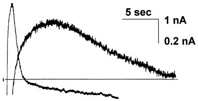
Figure 5.
Comparison of two outward K+ currents evoked by intracellular Ca2+ injection. Recordings were made in a single acutely isolated adult rabbit nodose neuron. A slow outward current (IK-slow) was activated by a 5-nA, 1-s iontophoretic Ca2+ injection at a holding potential of −50 mV. A second outward current (IK-medium) was activated at −20 mV (5 nA, 0.5 sec). IK-medium activates and decays completely before IK-slow reaches peak amplitude. IK-medium was blocked by 10 mM tetraethylammonium; IK-slow was blocked by 100 nM prostaglandin D2. The iontophoretic pipette was filled with a 0.2 M CaCl2 solution. Voltage-clamp currents were recorded with a second intracellular pipette. The discontinuous (switched) current injection mode of an Axoclamp II amplifier was used for both currentand voltage-clamp applications. The larger calibration value is for IK-medium. Population data is shown in Table 2.

