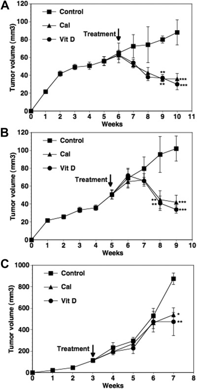
Fig. 1.
Effect of calcitriol and dietary vitamin D3 on the growth of xenograft tumors. MCF-7 xenografts were established in the flanks of intact female nude mice (premenopausal model). After 6 wk of tumor growth, the mice were given a vitamin D3-supplemented diet (Vit D) or basal diet + ip injections of calcitriol (0.05 μg/mouse) three times a week (Cal) for the following 4 wk. Mice in the control group (Control) were on the basal diet and received ip injections of the vehicle during this time period. Tumor volumes were measured weekly and calculated as described in Materials and Methods (A). In separate experiments, MCF-7 xenografts were established in the right and left fourth mammary fat pads of OVX nude mice (postmenopausal model) implanted with 60-d time-release androstenedione pellets and allowed to grow for 6 wk. The treatment groups were the same as described in A except that calcitriol was used at 0.025 μg/mouse. Tumor volumes in the experimental groups over the next 4 wk are shown (B). PC-3 xenografts were established in the flanks of intact male nude mice. After 3 wk of tumor growth, treatments were initiated as described in A except that calcitriol was used at 0.1 μg/mouse. Tumor volumes in the experimental groups over the next 4 wk are shown (C). Values represent mean ± se. The number of mice in the various experimental groups were 12–15 in the premenopausal and seven to nine in the postmenopausal models of BCa and six to eight in the PCa xenograft model. *, P < 0.05, **, P < 0.01 and ***, P < 0.001 compared with the corresponding control values.

