Figure 1.
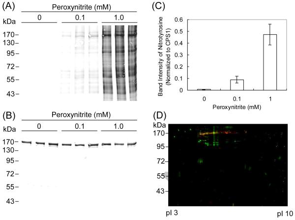
Western blot analysis of mouse mitochondrial proteins after treatment with peroxynitrite. (A) The mitochondrial fractions collected from three mice were individually incubated with each concentration of peroxynitrite (0, 0.1 and 1.0 mM) at 37°C for 10 min and subjected to 1D SDS gel electrophoresis. The blots were probed with antibodies to nitrotyrosine. (B) The blots were probed with antibodies to CPS1. (C) Densitometry analysis of nitration to CPS1 was performed using the Odyssey Infrared Imaging System. (D) The mitochondrial proteins pooled from three mice were incubated with 1.0 mM peroxynitrite at 37°C for 10 min and subjected to 2D gel electrophoresis. The blots of nitrated proteins (green) were superimposed with those of CPS1 (red). The overlap is represented by yellow.

