Figure 1.
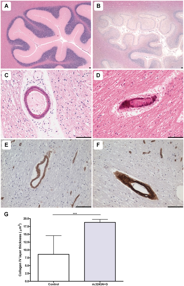
Pathological changes in small vessel morphology are observed in the cerebellum. Relative to control (A; haematoxylin and eosin), the cerebellar cortex of a patient harbouring m.3243A>G shows evidence of an ischaemic-like lesion involving the molecular, Purkinje cell and granular cell layers and the underlying white matter (B; Patient 6; haematoxylin and eosin). Arteriole mineralization is prominent in patients harbouring the m.3243A>G mutation (C and D; Patients 6 and 7; haematoxylin and eosin). Relative to control vessels (E; collagen IV), numerous patient vessels show increased deposition of collagen IV (F; Patient 7; collagen IV). Quantitation of the collagen IV immunopositive layer reveals a significant increase in collagen IV within the basement membrane of arterioles from patients with the m.3243A>G mutation relative to control vessels (G; ***P < 0.0001). Scale bars = 100 µm.

