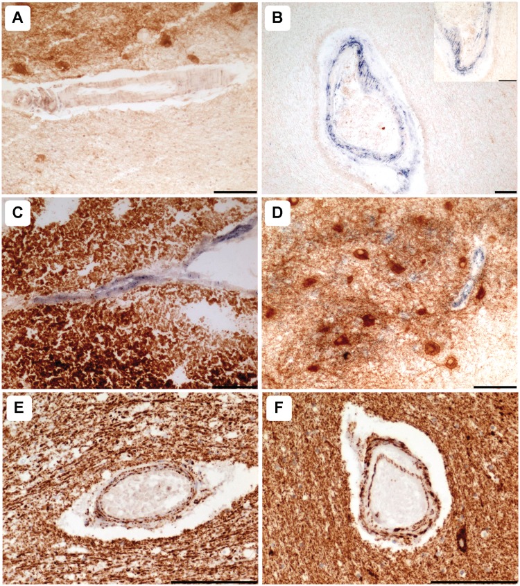
Figure 2.
Mitochondrial dysfunction is prominent in patients harbouring m.3243A>G and m.8344A>G point mutations. Relative to control vessels (A), COX-deficiency within the smooth muscle and endothelial cell layers was evident in cerebellar white and grey matter arterioles of patients with the m.3243A>G (B; Patient 7; COX-succinate dehydrogenase) and the m.8344A>G (C; Patient 9, COX-succinate dehydrogenase) mutations. Typically, capillary COX-deficiency was marked relative to dentate neurons where COX activity was intact (D; Patient 7, COX-succinate dehydrogenase). Relative to white matter vessels in controls (E), mitochondrial density was increased in many vessels of the patients with mitochondrial DNA disease (F, Patient 5, Porin immunohistochemistry). Scale bars = 100 µm.

