Figure 2.
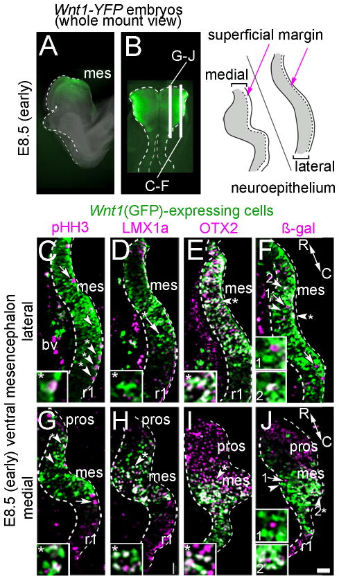
Molecular identity of Wnt1-expressing progenitors in early vMes. A–B: Whole mount E8.5 embryos indicate the location of sections that are shown. A: sagittal view. B: dorsal view. Illustration shows region of analysis. C–J: Sections from E8.5 Wnt1-Venus;TOPGAL embryo showing Wnt1(GFP)+ progenitors (green) in the mes and the indicated markers (magenta); note that Wnt1(GFP) was not expressed in rhombomere 1 (r1) or in the prosencephalon (pros). White arrowheads with asterisk show co-localization of Wnt1(GFP)+ progenitors and marker; white arrowheads show examples of Wnt1(GFP)+ only cells; white arrows show cells expressing only the marker. C,G: Wnt1(GFP)+ progenitors that were mitotic (pHH3+) were located in mitotic zone at the periphery of the tissue. D,H: LMX1a was co-localized with medial, but not lateral Wnt1(GFP)+ progenitors. E,I: OTX2 was co-localized with Wnt1(GFP)+ progenitors both medially and laterally; in medial sections the OTX2+/Wnt1(GFP)− prosencephalon (red) was apparent. F,J: There were Wnt1(GFP)+ cells that responded to canonical WNT signaling in the medial domain as evident by overlap with β-gal from the TOPGAL reporter (Fig 2F). However, fewer Wnt1(GFP)+ cells responded to canonical WNT signaling in the lateral domain (Fig. 2J). Scale bar = 32μm (C–J).

