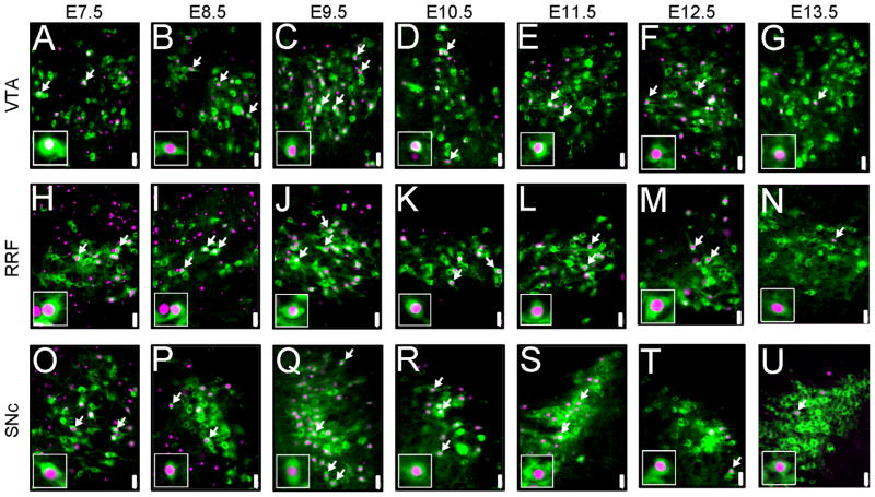
Figure 6.
MbDA neurons in the VTA, RRF, and SNc domains were derived from Wnt1-expressing progenitors over a prolonged time period. MbDA neurons in adult Wnt1-CreERT;TaumGFP mice marked by tamoxifen administration at distinct 24 hour time points between E7.5–E13.5. A–G: VTA (medial) MbDA neurons. H–N: A8/RRF (retrorubral field) MbDA neurons. O–V: SNc (lateral) MbDA neurons. MbDA neurons are indicated by TH immunolabeling (green) and the Wnt1 lineage is indicated by nuclear β-gal immunolabeling (red). Arrows show examples of neurons that expressed both TH and β-gal. Scale Bar = 32μm.

