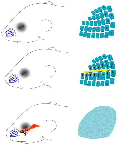
Figure 2.
Schematic diagram illustrating the classical structural plasticity in the barrel cortex following row C whisker lesions or infraorbital nerve transection. These effects are only seen when peripheral lesions are performed up to postnatal day 3. The patterns and deficits are routinely assessed by histochemical stains such as succinic dehydrogenase or cytochrome oxidase histochemistry or with immunohistochemistry for TCA markers such as 5-HTT or vesicular glutamate transporter 2 or by Nissl or Golgi stains for neuronal and dendritic organization.

