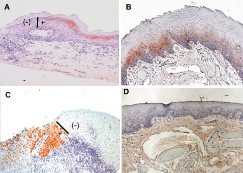
Figure 3. SIBLING/MMP immunoreactivity at histologically negative resection margins of OSCC.
(A) Example of DSPP expression scored as “1+” indicating >10% <50% of cells at resection margin showing immunoreactivity with DSPP (monoclonal antibody, LF-Mb21). (B) Expression scored as “2+” where 50% <75% of cells at histologically negative margins show immunoreactivity to BSP (antibody, LF-Mb25). (C) Expression scored as “3+” with more than 75% of normal epithelial cells at resection margins positive with MMP-9 (antibody LF-184). (D) Representative non-immune control showed expression scored as 0 for no immunoreactivity at histologically negative margin. Chromogenic staining (reddish-brown color) was achieved with 3-amino-9-ethylcarbazole (AEC) and counterstained with hematoxylin. Bars (I) in A and C show point of abrupt end of DSPP and MMP-9 expression, respectively, at the margins demarcating area of histologically negative but DSPP/MMP-9 positive (+) from histologically negative and DSPP/MMP-9 negative (-) portions of the margin.

