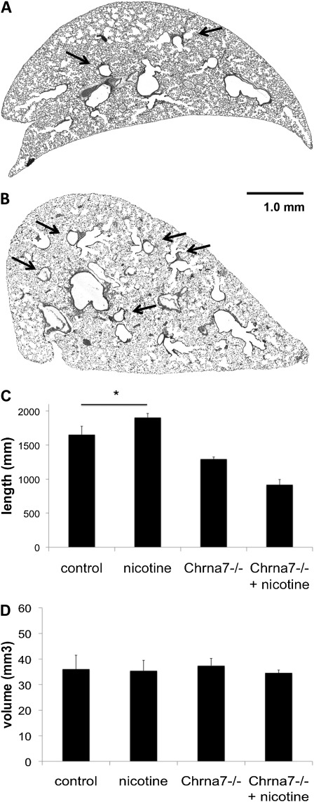
Figure 3.
Adult mice exposed to nicotine prenatally have greater airway length without a concomitant change in volume. C57BL6/J female mice were administered either untreated water or water containing 100 μg/ml nicotine before timed breeding and through gestation. Lungs were obtained from age-matched offspring at 8 weeks of age. Unexposed lungs (A) have less airway branching (arrows highlight selected airways) compared with lungs exposed to nicotine in the prenatal period (B) on gross examination. Using standard stereologic techniques, total airway length and volumes were estimated. Mice exposed to prenatal nicotine have greater total airway length (C) without any change in volume (D) when compared with unexposed wild-type control animals. Animals deficient in α7 nAChR (Chrna7−/−) exposed to prenatal nicotine do not demonstrate greater total airway length when compared with unexposed animals deficient in α7 nAChR (C) (n = 3–8 per group). Error bars denote SD (*P < 0.05 compared with control). Scale bar, 1 mm.

