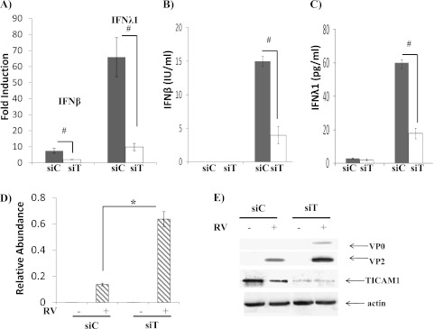
Figure 5.
TICAM1 was required for IFN production and epithelial anti-RV response. RV-induced IFN expressions were dependent on TICAM1. Control siRNA (siC) and TICAM1 siRNA (siT) were transfected into NCI-H292 cells. Twenty-four hours later, NCIH292 cells were infected with RV16 at MOI = 10 for 24 hours. RV was then thoroughly washed out as described elsewhere (21). Total cellular RNA, protein, and media were collected 24 hours later. (A) IFN-β and IFN-λ1 expressions were determined by real-time PCR. The data are presented as the fold inductions comparing infected RV with mock (saline)-infected control cells. #P < 0.05 when comparing siC and siT (n = 5). (B) ELISA was used to determine the concentration of IFN-β in culture media. The data are presented as IU/ml. #P < 0.05 (n = 5). (C) ELISA was used to determine the concentration of IFN-λ1 in culture media. The data are presented as pg/ml. #P < 0.05 (n = 5). (D) RV-positive strand RNA was determined by real-time PCR. The data are presented as the relative abundance due to the lack of RV in mock-infected cells as described. #P < 0.05 when siC-transfected cells were compared with siT-transfected cells (n = 5). (E) Western blot analysis was used to determine TICAM1 and RV coat proteins (VP0 and VP2) as described elsewhere (21). All images are representative of at least three independent experiments.

