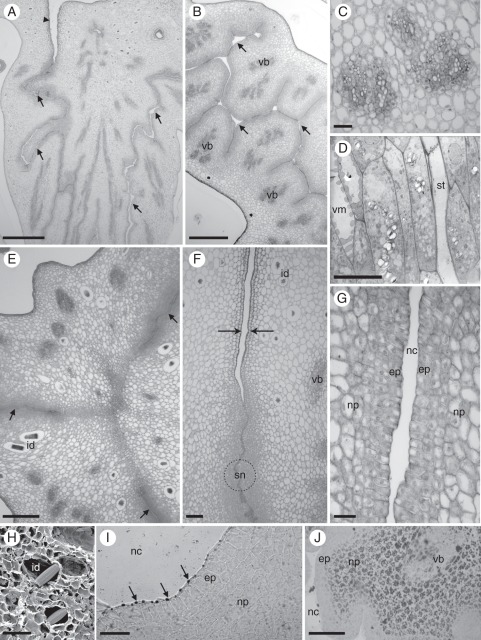
Fig. 2.
Septal nectary structure of Ananas ananassoides. (A) Ovary longitudinal section showing septal nectary (arrows) and apical orifice (arrowhead). Scale bar = 500 µm. (B) Cross-section of the middle region of the ovary showing the nectar-secreting channel (arrows). vb, vascular bundle. Scale bar = 300 µm. (C) Detail of (B) showing vascular bundles. Scale bar = 150 µm. (D) TEM image of the vascular bundle showing vessel member (vm) and sieve tube member (st); note large amyloplasts in the parenchyma cells. Scale bar = 5 µm. (E) Cross-section of the ovary at the ovule attachment region showing the nectar-secreting channels (arrows) with a linear outline; note idioblasts with raphides (id). Scale bar = 200 µm. (F) Part of the ovary apical region showing a progressive lack of secretory tissue. Note the epidermis with cuticle (arrows), septal nectary (sn) and idioblasts (id) with raphides. Scale bar = 50 µm. (G) Part of the septal nectary in cross-section with multiseriate epithelium (ep), nectary parenchyma (np) and nectar-secreting channel (nc). Scale bar = 50 µm. (H) SEM image of the ovary showing idioblasts (id) with raphides. Scale bar = 20 µm. (I) Section of an NADI-stained nectary showing reaction products (arrows) between the protoplast and the cell wall of epithelial cells. Scale bar = 50 µm. (J) Distribution of starch grains in the septal nectary treated with Lugol's reagent. ep, epithelium; np, nectary parenchyma; vb, vascular bundle. Scale bar = 100 µm.

