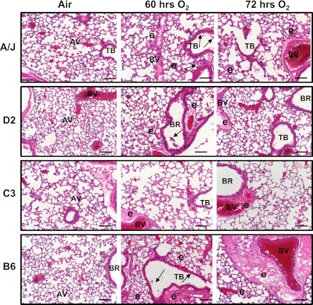
Figure 3.
Differential pulmonary histopathology developed after hyperoxia exposure in four inbred strains of mice. Representative light photomicrographs of left lung sections from A/J, DBA/2J (D2), C3H/HeJ (C3), and C57BL/6J (B6) mice (n = 3–5/group) of air controls or hyperoxia (60 and 72 h) stained with H and E. Arrows indicate epithelial cell sloughing. AV, alveoli; BR, bronchi or bronchiole; BV, blood vessel; e, alveolar and tissue (perivascular-peribronchiolar) edema; and TB, terminal bronchiole. Scale bars, 100 μm.

