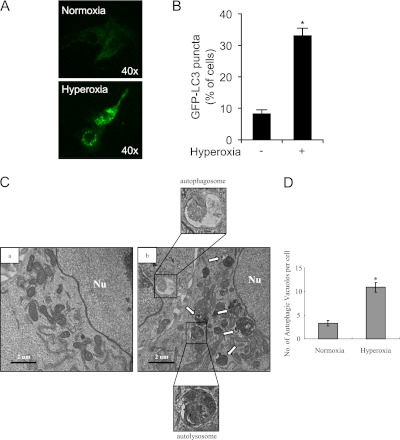
Figure 2.
Hyperoxia induces autophagy in cultured epithelial cells. Beas-2B cells were transfected with 20 μg of GFP-LC3B using Lipofectamine TM2000 (Invitrogen, Carlsbad, CA) in OPTI-MEM and then exposed to normoxia or hyperoxia for 24 hours. The cells were observed under fluorescent microscopy. (A) Representative image is shown (original magnification: ×40). (B) Percentages of cells with punctuated GFP-LC3 were quantified. Results are expressed as means ± SEM of more than 100 cells per sample. Similar results were obtained from three independent experiments. *P < 0.01 compared with control. (C) The representative electron micrograph shows the ultrastructure of Beas-2B cells exposed to standard cultured conditions (normoxia) or hyperoxia for 24 hours. Representative morphology of autophagic vacuoles is shown, with evidence of autophagosome formation. Nu = nucleus; white arrows, autophagic vacuoles. Scale bar, 2 μM. (D) The average number of autophagic vacuoles was determined from randomly selected samples of more than 30 cells. The total numbers of autophagosomes per cell are expressed as means ± SEM. *P < 0.01 compared with control.

