Figure 3.
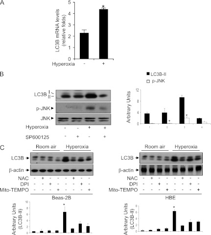
Involvement of c-Jun N-terminal kinase (JNK) in hyperoxia-induced autophagy. (A) Beas2B cells were exposed to normoxia and hyperoxia (12 h). mRNA was isolated and reverse transcribed, and Taqman real-time PCR was performed using LC3B primer and probe. Relative folds of increased mRNA transcription were displayed. (B) Total protein extracts (25 μg) were obtained from Beas-2B cells exposed to hyperoxia for 12 hours in the presence or absence of SP600125 (5 μM) and were subjected to immunoblot analysis with antibodies against LC3B, JNK, and phospho-JNK (pJNK). (C) Beas-2B cells or HBE cells were exposed to normoxia or hyperoxia for 24 hours in the presence or absence of N-acetyl-L-cysteine (NAC) (5 mM), diphenyleneiodonium (DPI) (10 μM), and mito-TEMPO (100 μM). Immunoblot analyses were performed to determine the levels of LC3B, and β-actin served as the standard. All results represent three independent experiments.

