Figure 4.
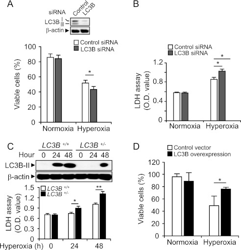
Cellular function of LC3B in hyperoxia-induced cell death. (A) Beas-2B cells were transfected with control-siRNA and LC3B-siRNA for 30 hours followed by exposure to normoxia or hyperoxia for 48 hours. Cells were labeled with FITC-Annexin V and propidium iodide (PI) and analyzed with flow cytometry (n = 7). Annexin V(−) and PI(−) cells were defined as live cells. The expression of LC3B was determined by Western immunoblot analysis. β-actin served as the standard (inset). (B) Cells were treated as in (A). Lactate dehydrogenase (LDH) levels in the supernatant were measured by Cytotoxicity Detection Kit (Roche Applied Science, Indianapolis, IN) (n = 8). Results are expressed as means ± SEM of indicated numbers of independent experiments. *P < 0.05 compared with indicated groups. Statistical significance was determined by ANOVA with multiple comparisons. (C) Primary lung fibroblasts were harvested from wild-type and LC3B+/− mice. Equal numbers of cells (1 × 10⁶ cells per dish) were cultured on the 60-mm dishes and exposed to hyperoxia. Cells were harvested at 24 and 48 hours. (Upper panel) Total protein extracts (25 μg) were subjected to Western immunoblot analysis with antibodies against LC3B. (Lower panel) LDH levels in the supernatant were measured by Cytotoxicity Detection Kit. Results are expressed as means ± SEM of five independent experiments. *P < 0.05 and **P < 0.03 compared with indicated group. (D) Beas2B cells were transfected with control vector and LC3B overexpression clones. After 24 to 30 hours, cells were exposed to hyperoxia. After 48 hours, cell viability was determined. Results are expressed as means ± SEM of three independent experiments. *P < 0.05.

