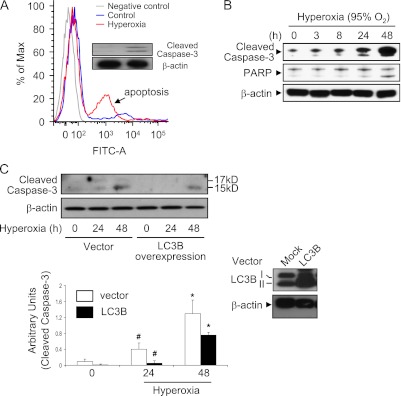
Figure 5.
Effect of LC3B in hyperoxia-induced epithelial cell apoptosis. (A) Primary mouse lung epithelial cells were isolated from C57/BL6 mice. Cells were then exposed to room air (RA) or hyperoxia (24 h). Annexin V and PI were used to stain cells, and fluorescence-activated cell sorter analysis was performed. Inset: Cleaved caspase 3 was determined using Western blot analysis. (B) Beas-2B cells were exposed to hyperoxia for up to 48 hours and harvested at the indicated times. Total protein extracts (25 μg) were subjected to immunoblot analysis with antibodies against the cleaved form of caspase-3 (c-caspase-3) or poly-(ADP ribose) polymerase (PARP). β-actin served as the standard. (C) Beas-2B cells were transfected with control vectors or LC3B overexpression clones. After 24 hours, cells were exposed to hyperoxia (24 and 48 h). Hyperoxia-induced cleaved caspase 3 (active form) was determined using Western blot analysis. Overexpression of LC3B was confirmed by protein level determined using Western blot analysis (inset). All results represent three independent experiments.

