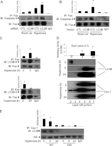
Figure 6.
Crosstalk of autophagy with apoptosis. (A) Beas-2B cells were transfected with control siRNA or LC3B siRNA. After 24 hours, cells were exposed to hyperoxia (4 h). Co-IP assays were performed to detect the interaction between Fas and caspase 8. (B) Beas-2B cells were transfected with control vector or LC3B expression vector. After 24 hours of transfection, cells were exposed to hyperoxia (4 h). Co-IP assays were performed to detect the interaction between Fas and caspase 8. (C) Beas2B cells were exposed to normoxia or hyperoxia for 4 hours (upper panels) and 24 hours (lower panels). The interaction between Fas and LC3B was determined by co-IP assays. (D) Beas2B cells were exposed to normoxia or hyperoxia for 4 hours. Lipid rafts were isolated, and the amount of LC3B trafficking into the lipid raft fraction was determined using Western blot analysis. (E) Beas2B cells were transfected with caveolin-1 wild-type Y14 clone and Y14D (tyrosine to aspartate) mutant clones. Cells were then exposed to hyperoxia (24 or 48 h). LC3B and cav-1 interaction was determined using co-IP assays and Western blot analysis. NS = nonspecific binding. Results are representative of three independent experiments.

