Figure 2.
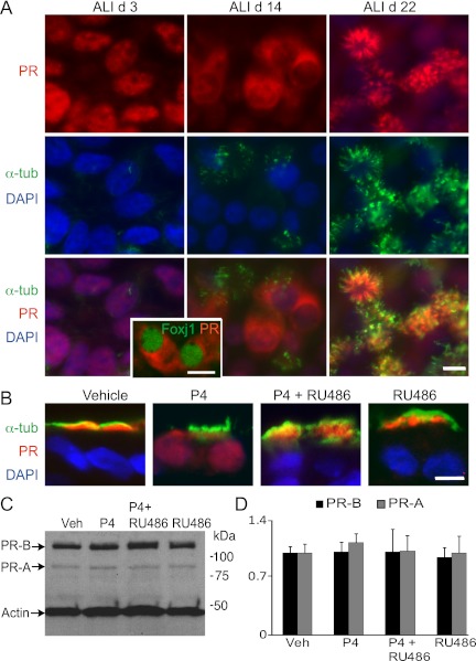
PR localization is differentiation and ligand dependent in airway epithelial cells. (A) Differentiation-dependent PR expression of hTEC preparations immunostained for PR (red) and acetylated α-tubulin (green) shown en face. Nuclei are stained with DAPI (blue). ALI Day 3 shows PR in the nuclei of all cells. As differentiation proceeds, PR moves to the cytoplasm in some cells (ALI Day 14). Inset shows hTECs immunostained for PR (red) and Foxj1 (green) during differentiation, demonstrating cytoplasmic PR during early ciliogenesis (ALI Day 11). In differentiated hTECs, PR is expressed in the cilia (ALI Day 22). Top right panel shows PR expression in cilia, and top left panel shows PR expression in nuclei of nonciliated cells, within a plane of focus below the level of the cilia. (B) P4-induced nuclear localization of PR in fully differentiated hTEC preparations. hTECs treated with P4, mifepristone (Mife), or P4 plus mifepristone, and immunostained as in (A). (C) Protein blot analysis of hTECs treated as in (B) show no change in the expression of PR isoforms. (D) Densitometry analysis of studies in (C) expressed as the mean (±SD) of fold change of PR-A and -B normalized to actin and relative to vehicle-treated samples from three independent experiments. Scale bars, 10 μm in (A and B).

