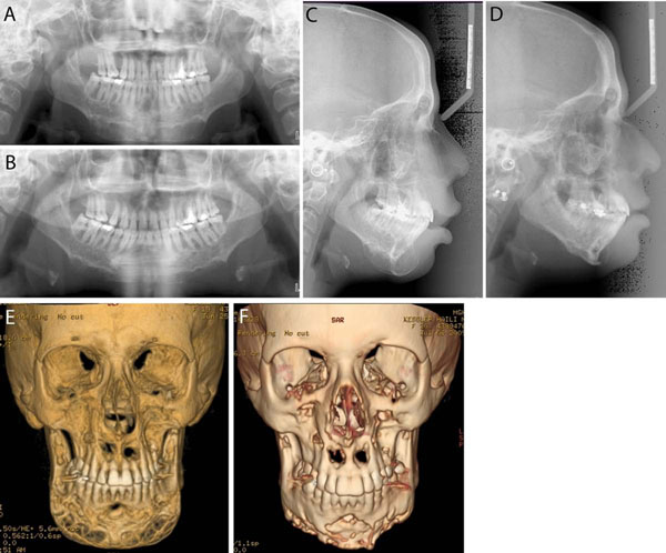
Figure 4.
Imaging studies of the patient in Figure 3 pre-treatment (A, C, E) and post-treatment (B, D, F). A and B. Panoramic radiographs. C and D. Lateral cephalograms. E and F. 3D CT scans. Pre- and post-treatment images show improvement of the giant cell lesions and the contours of the mandible.

