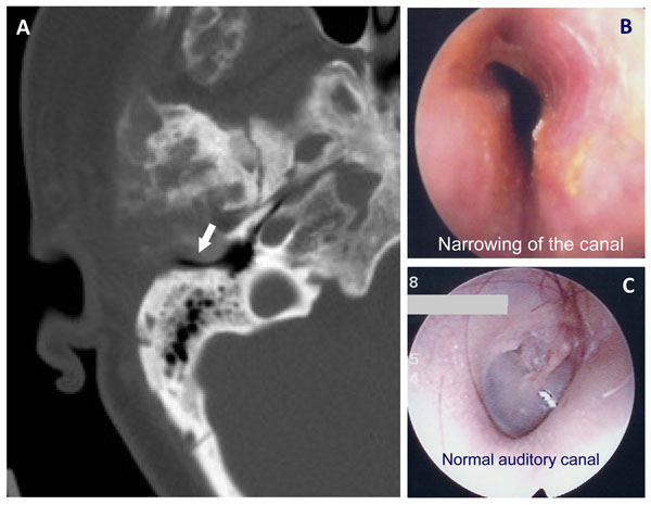
Figure 11.
Narrowing of the external auditory canal due to fibrous dysplasia (FD). A) A CT image of a coronal slice through the temporal bone shows a narrowed external auditory canal (arrow) B) Narrowing of the canal is shown and can be compared to a normal canal in (C). The arrow on the CT image (A) demonstrates narrowing of the canal. This has resulted in hearing loss. The clinical images on the right compare a canal narrowed by FD to a normal external auditory canal.

