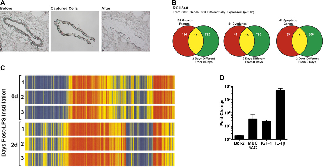
FIGURE 1. Microarray analysis of laser-capture microdissected (LCM) rat airway epithelia.
(A) Light micrographs of the rat large airways before and after laser-capture microdissection of epithelial cells. (B) Venn-diagram clustering of genes expression pattern obtained by microarray analysis of LCM-captured epithelial cells from LPS-instilled and control rats. (C) Heat-map displaying differential gene expression pattern (p<0.05) of microdissected airway epithelia mRNA from rats instilled with LPS or non-instilled controls (n=3 rats per group). (D) Relative quantities of Bcl-2, MUC5AC, IGF-1, and IL-1β mRNA levels in LCM-captured epithelial cells from LPS-instilled rats compared to control rats as quantified by qRT-PCR.

