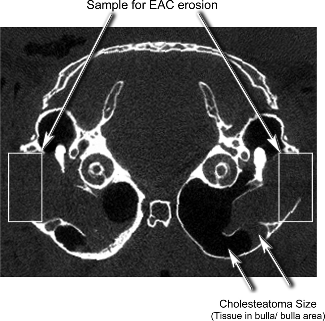
Figure 3.
An axial microCT scan of a gerbil head with induced cholesteatoma demonstrating morphometric measures. The area of bone resorption was determined by the length of remaining bone in a standardized box. The area occupied by the cholesteatoma was determined by the area of soft tissue (radio-opaque) divided by the area of soft tissue plus area of gas (radiolucent)

