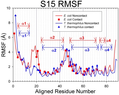
Figure 4. RMSF values for S15.
E. coli proteins are represented in red with squares indicating contact residues and T. thermophilus proteins are colored blue with triangles for contacts. In these figures, the proteins have been sequentially aligned to demonstrate the behaviors of the conserved structural elements. Aligned Residue Numbers, therefore, do not necessarily reflect the actual residue indices of the protein sequence.

