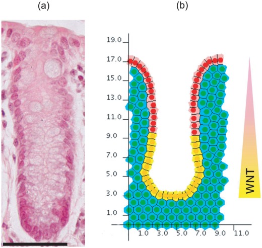
Figure 13. The cross-sectional crypt configuration.
(a) A haematoxylin and eosin stained wax section of a murine colonic crypt. Scale bar =  . (b) The cross-sectional model geometry, plotted against the axes used to mark cell
  -coordinates for the results presented. A linearly decreasing gradient of Wnt factors is imposed in the cross-sectional model, which is normalised to 1 at the base of the crypt, and 0 at the crypt collar. A threshold is prescribed such that cells in the region of insufficient Wnt factors are defined to be terminally differentiated. Thus, proliferating and non-proliferating epithelial cells are indicated in yellow and pink respectively, whilst non-proliferating stromal cells are indicated in green.

