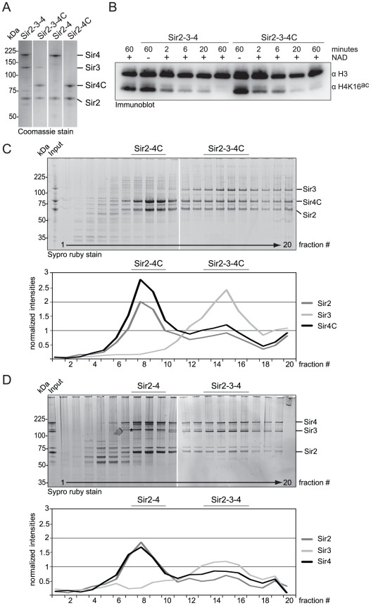
Figure 4. Sir4C can form a stable and active SIR complex in a recombinant system.
A) SIR complexes as indicated were purified form co-infected insect cells. 1 µg of each complex was run on a SDS-PAGE and visualized by Coomassie staining. B) Purified Sir2–Sir3–Sir4 and Sir2–Sir3–Sir4C complex were incubated with histone octamers acetylated at H4K16 with or without the essential cofactor NAD. The deacetylation reaction was stopped after various time points by the addition of sample buffer and monitored by immuno blotting for H4K16ac and H3, for equal loading. C, D) Sir2–Sir3–Sir4 or Sir2–Sir3–Sir4C complexes were analyzed by density gradient sedimentation. Fractions were run on 4–12% NuPAGEs Novex Bis-Tris Gels and stained with Sypro Ruby dye. Intensities of Sir2, Sir3 and Sir4 full length proteins were quantified (QuantityONE) and plotted in line graphs. The asterisk in D) indicates a Sir4 degradation band that runs very closely to Sir3.

