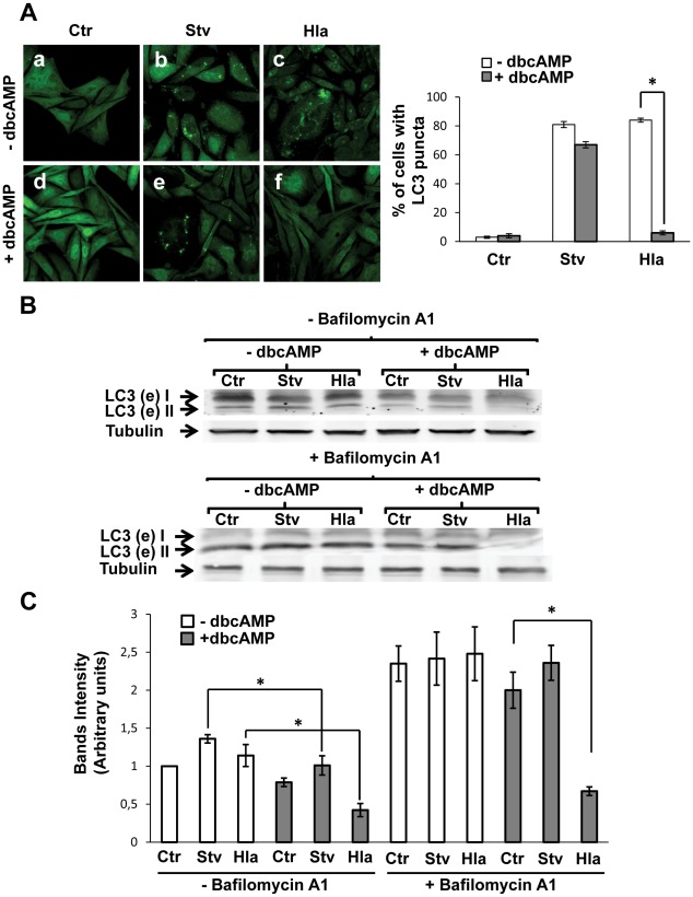
Figure 1. cAMP inhibits Hla-induced autophagy.
(A) GFP-LC3 CHO cells were preincubated with 1 mM dybutiryl cAMP (+dbcAMP, panels d, e, and f) for 30 min and then were treated for 4 h with 10 µg/ml of α-hemolysin (Hla) or subjected to starvation conditions (Stv) in the continuous presence of dbcAMP. Cells without any treatment were used as control (−dbcAMP, panels a, b, and c). Cells were immediately analyzed by confocal microscopy. A quantification of the percentage of cells presenting LC3 puncta (i.e., stimulated cells) upon incubation with the different conditions is shown in the right panel (n = 100 cells/condition, * p<0.05 paired Student's t-test). These data are representative of three independent experiments. (B) GFP-LC3 CHO cells were incubated with complete medium in the absence (−dbcAMP) or presence of dbcAMP (+dbcAMP) and treated for 4 h with 10 µg/ml of α-hemolysin (Hla) or subjected to starvation conditions (Stv) with (lower panel) or without (upper panel) bafilomycin A1 to block lysosomal degradation. Afterwards, cells were lysed with sample buffer and the samples were subjected to Western blot analysis using a rabbit anti-LC3 and the corresponding HRP-labeled secondary antibody, and subsequently developed with an enhanced chemiluminescence detection kit. These data are representative of three independent experiments. (C) The band intensities of two independent experiments were quantificated with the Adobe Photoshop program, and normalized against tubulin. * p<0.05 (paired Student's t-test).

