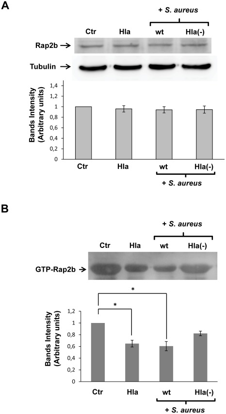
Figure 11. Active Rap2b is decreased to allow the autophagy response induced by Hla and S. aureus.
(A) HeLa cells were incubated for 4 h with complete medium in the presence (Hla) or absence (Ctr) of 10 µg/ml α-hemolysin or they were infected for 4 h with the wt strain of S. aureus (wt) or the α-hemolysin deficient mutant (Hla−). Afterwards, cells were lysed with sample buffer and the samples were subjected to Western blot analysis using a rabbit anti-Rap2b and the corresponding HRP-labeled secondary antibody. The bands were subsequently developed with an enhanced chemiluminescence detection kit. A quantification of the bands intensities with the Adobe Photoshop program is shown in the lower panel. These data are representative of two independent experiments. (B) HeLa cells were incubated for 4 h with complete medium in the presence (Hla) or absence (Ctr) of 10 µg/ml α-hemolysin or they were infected for 4 h with the wt strain of S. aureus (wt) or the α-hemolysin deficient mutant (Hla−). Cells were disrupted and whole cell lysates were subjected to pull-down assays using GST-Ral-GDS-RBD-sepharose. The levels of GTP-bound Rap2b were determined as was described in Materials and Methods by Western blot analysis using a rabbit anti-Rap2b and the corresponding HRP-labeled secondary antibody. The bands were subsequently developed with an enhanced chemiluminescence detection kit. The band intensities were quantificated with the Adobe Photoshop program is shown in the lower panel. * p<0.05 (paired Student's t-test). These data are representative of two independent experiments.

