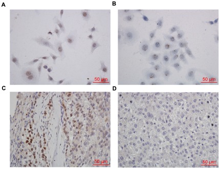
Figure 6. These photomicrographs show in situ hybridization results for human papillomavirus positive hepatocellular carcinoma.
(A) Hep G2 cells were with the punctate signal pattern of HPV DNA. (B) HeLa cells were served as positive control. (C) Hepatocellular carcinoma was with diffuse signal pattern of HPV staining. (D) No signal was found in hepatoma carcinoma cells of this HPV- negative specimen.

