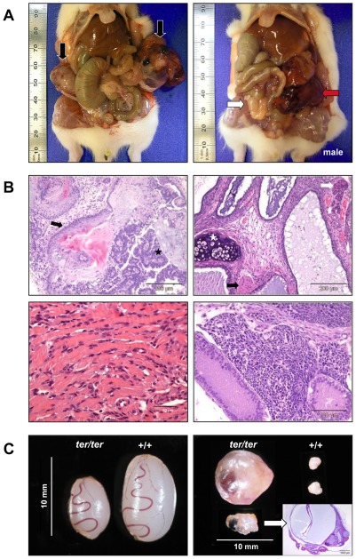
Figure 1. Teratomas and gonads in ter/ter rats.
(A) Left: Bilateral GCTs in a 6 week old female rat (black arrows: OGCTs). Right: Unilateral GCT in male rat (white arrow: non-neoplastic testes; red arrow: left TGCT). (B) HE staining of teratomas. Top left: ectodermal tissues in the ovarian teratoma of a 5-week-old female; neural tube formation (black star) and squamous epithelium (black arrow). Top right: mesodermal and endodermal tissues in the ovarian teratoma of a 9-week-old female; cartilage (white star), glandular structures (white arrow) and skeletal muscle (black arrow). Bottom left: heart muscle-like tissue from a contractile ovarian teratoma. Bottom right: immature neuronal tissue from a testicular teratoma of a 6-week-old male. (C) Rat gonads from 3-week-old siblings. Left: non-tumorous ter/ter testes are significantly smaller than wild type. Right: physiological wild type ovaries compared to an ovarian teratoma and an abnormal, cystic ovary (confirmed by histological section and HE staining).

