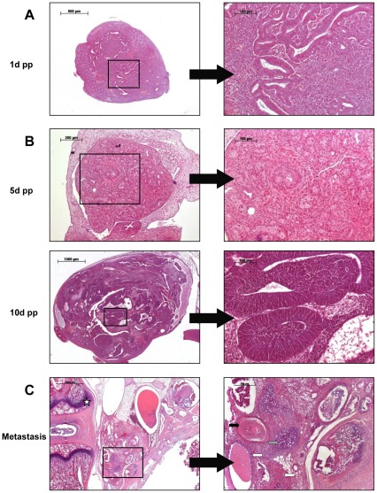
Figure 4. HE staining of early tumorigenesis and metastasis.
(A) Male. Tumor formation in the testis of a 1-day-old Dnd1-deficient male. 5× and 40× (B) Female. Neoplastic transformation in the ovary of ter/ter females at 5 and 10 days post partum. Top: 10× and 40×. Bottom: 2.5× and 40×. (C) Metastasis. Teratoma in adjunction to the vertebral column (white star). Tissue of ectodermal (squamous epithelium, black arrow), mesodermal (cartilage, gray arrow) and endodermal (glandular structures, white arrow) origin was identified. 2.5× and 10×.

