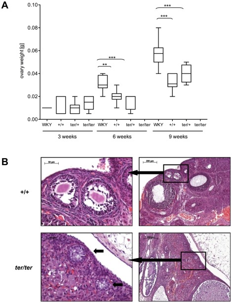
Figure 7. Non-tumorous ovaries.
(A) Box plot analysis of non-tumorous ovaries from WKY/Ztm and WKY-Dnd1ter/Ztm with wild type (+/+), heterozygous (ter/+) and homozygous (ter/ter) genotype (**: p<0.01; ***: p<0.001). (B) HE staining of wild type (+/+, top panel) and mutant ovaries (ter/ter, bottom panel) of WKY-Dnd1ter/Ztm rats at 6 weeks of age. While different stages of follicle maturation (top panel, left) and primary follicles with a central oocyte (top panel, right) were evident in the wild type ovary, few follicle-like structures without germ cells (bottom panel, right, arrows) remained in the ter/ter ovary.

