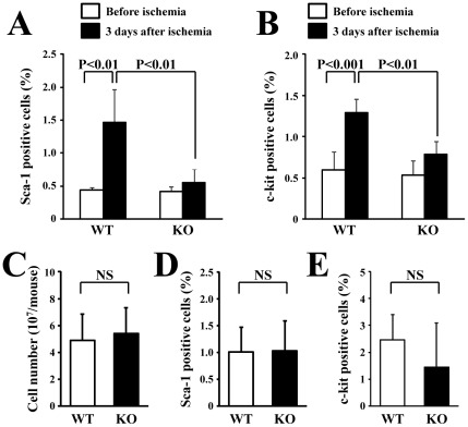
Figure 3. Mobilization of BM-derived cells in the peripheral blood.
Flow cytometric analysis showed that the mobilization of BM-derived Sca-1- (A) and c-kit-positive cells (B) in the peripheral blood significantly increased in the WT mice but not in the HSF1-KO mice, 3 days after ischemia (n = 4 animals/group). However, the total amount (C) or the subpopulation of Sca-1- (D) and c-kit-positive cells (E) in BM cells did not differ between WT and HSF1-KO mice (n = 3 animals/group).

