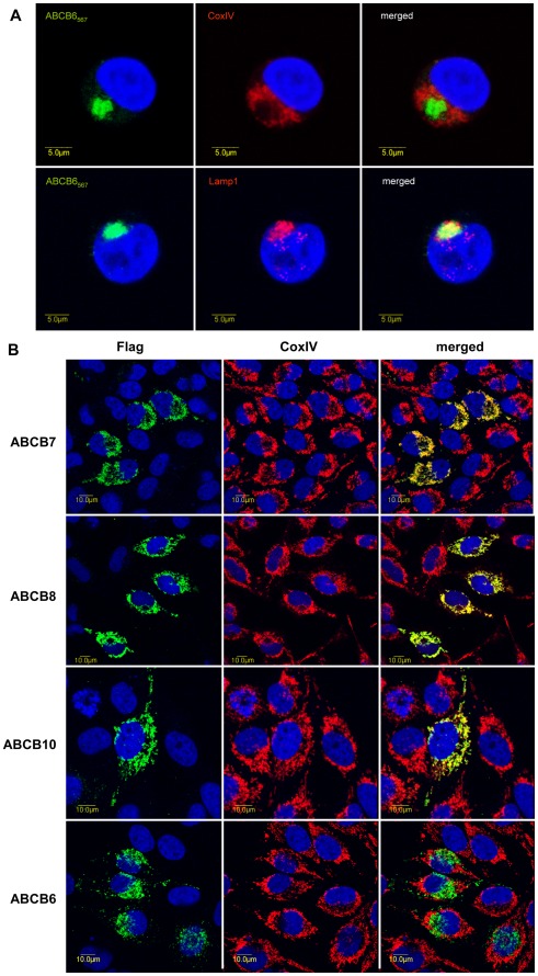
Figure 2. Determination of the subcellular localization of ABCB6 by double immunofluorescence labeling and laser-scanning confocal microscopy. A.
. Expression of the endogenous ABCB6 protein in K562 cells was visualized by the monoclonal ABCB6-567 antibody (green), lysosomes and mitochondria were labeled with LAMP1 (red, lower panel) and CoxIV antibodies (red, upper panel), respectively. ABCB6 colocalizes with LAMP1 (yellow on the overlay, r = 0.79), but not with CoxIV (r = 0.14). B. Flag-tagged ABC proteins were expressed following transient transfection of HeLa cells. The cDNA-derived ABC transporters were visualized with an anti-FLAG tag antibody (green, left panel); mitochondria were labeled with CoxIV (red, middle panel). Whereas the canonical mitochondrial ABC transporters are confined to the mitochondria (merged image, right panel, r = 0.87, r = 0.7, r = 0.84 for ABCB7, ABCB8 and ABCB10, respectively), ABCB6 does not show colocalisation with the mitochondrial marker (merged image, right panel, r = 0.13).

