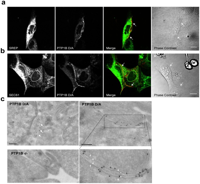
Figure 2. The endoplasmic reticulum lies in close proximity to the plasma membrane at regions of cell-cell contact.
(a) MDCK cells co-expressing PTP1B D/A-mCherry and a general marker for the endoplasmic reticulum, stress-related ER protein (SREP-YFP); or (b) PTP1B D/A-mCherry Sec61-YFP, a marker for the rough ER. Arrows indicate region of cell-cell contact. Scale bars correspond to 10 µm. (c) Immunogold electron microscopy shows PTP1B D/A localization in the ER (arrows). No significant labeling was detected in PTP1B-null cells (left bottom image). Significant immunogold labeling was also detected at regions of cell-cell contact (right image). The boxed area, which is magnified below, shows the region of cell-cell contact that reveals labeling at the ER (arrows) proximal to the PM (arrowheads). Also see Fig. S2. Scale bars correspond to 0.2 µm.

