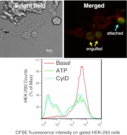
Fig. 2.
Phagocytosis of apoptotic HPB cells by HEK-293 cells transfected with P2X7: upper panel bright field image (left) and confocal image (right) showing a HEK-293 cell transfected with DsRed tagged P2X7 (red) containing two engulfed CFSE-labeled HPB cells and another which is tethered or attached to the HEK cell. Images were acquired by an Olympus IX81 confocal microscope (×600). Lower panel: a typical flow cytometry histogram showing phagocytosis of CFSE-labeled HPB cells by P2X7-DsRed transfected HEK-293 cells. HEK-293 cells were labeled with BODIPY 630/650-SE first and pre-treated with 1 mM ATP or 20 μM CytD for 15 min prior to incubation with HPB cells. BODIPY+/DsRed++ HEK-293 cells were gated for analysis. From Gu et al. [29]

