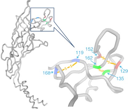
Fig. 5.
Predicted extracellular and transmembrane domains of human P2X7 structure. The P2X7 model was based on the crystal structure of zebrafish P2X4 in the closed state (Ref. [36]) using the online Protein Homology/analogY Recognition Engine (PHYRE, V2.0). The six conserved cysteine residues in the exposed “dolphin nose” region (residues 115–168) form three disulfide bonds shown between the matching colors

