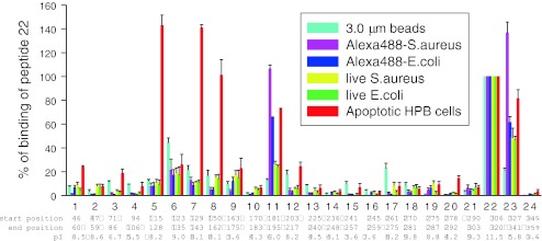
Fig. 6.
The binding of short peptides mimicking P2X7 extracellular domain sequences to a range of phagocytic targets. 24 biotin-tagged peptides were incubated at a concentration of 10 μg/mL with either YG beads (3 μm, 5 μL), Alexa 488 conjugated Staphylococcus aureus or Escherichia coli (2 mg/mL, 5 μL), live S. aureus or E. coli (OD690 = 0.9, 10 μL each) or apoptotic HPB cells (5 × 10⁶/mL, 200 μL) for 30 min, followed by washing and incubation with HRP-labeled streptavidin for 30 min. Particles were washed twice, and resuspended in 100 μL of HRP-labeled streptavidin (Jackson ImmunoResearch Lab, 1:2,000 diluted with PBS containing 1% BSA) for 30 min. After two washes, 500 μL of SuperSignal West Pico was added and the chemiluminescence was measured by a Glo 20/20 luminometer (Promega). From Gu et al. [29]

