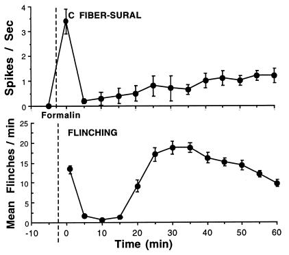
Figure 1.
(Upper) C fiber activity recorded in situ in rats from single sural nerve fibers, identified by their conduction velocity and modality as C fibers. Immediately after formalin injection (as indicated by the dashed line) into their receptive fields, high activity was observed in high-threshold C nociceptive afferent fibers (as well as in A beta and A delta fibers, data not shown). At later intervals, activity was observed in all mechanically sensitive C fibers, at rates that were less (1/2–2/3) than those achieved initially (adapted from ref. 6). (Lower) Frequency of flinching as measured by an automated motion detector is plotted at 5-min intervals after the injection of formalin into the paw at the time indicated by the vertical dashed line. As indicated, the flinching behavior displays a biphasic occurrence (phase 1 and phase 2). The data represent the mean ± SEM of eight rats.

