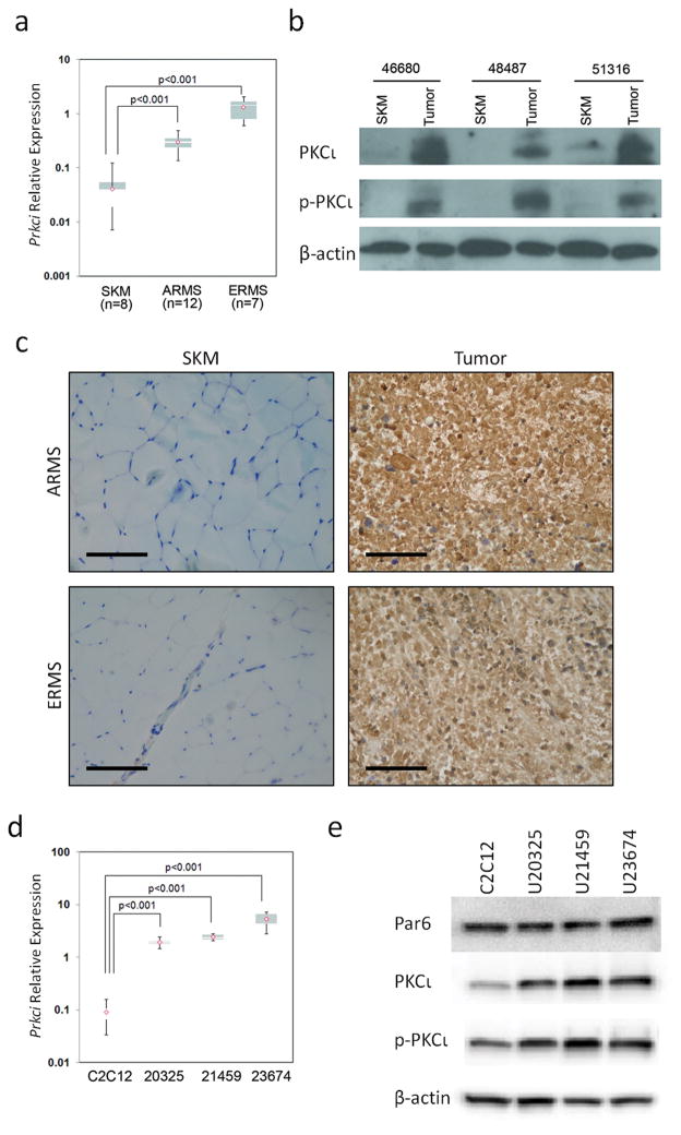
Figure 2.
Over-expression of PKCι in mouse ARMS. (a) Quantitative RT-PCR show Prkci is over-expressed in mouse ARMS and ERMS compared to normal skeletal muscle (7.5 and 33 fold respectively, p<0.001). (b) Immunoblotting for PKCι shows high expression and activation in mouse tumors compared to matched skeletal muscle. (c) PKCι immunohistochemistry of mouse tumors. Size bar = 0.05 mm. (d) Prkci is over-expressed in mouse ARMS primary cell cultures compared to mouse myoblast C2C12. (e) Immunoblots show Par6, PKCι and phospho-PKCι expression in mouse ARMS primary cell cultures. See Supplementary Figure S2 for full length blots of Figure 2e.

