
Figure 3. Lesion location encoding.
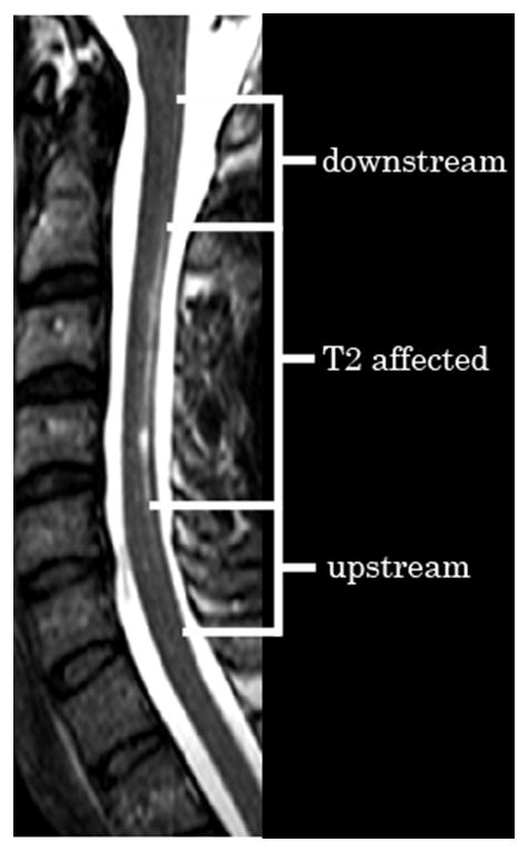
This axial T2 weighted image in a subject with NMO illustrates a longitudinally extensive lesion confined to the spinal cord dorsal white matter. Within the sensory posterior column, areas are labeled upstream of the lesion, T2 affected and downstream of the lesion.