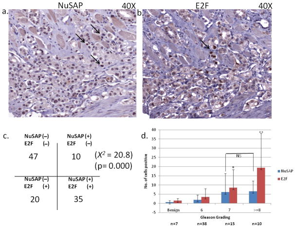
Figure 6.
Immunostaining of prostate TMA with NuSAP and E2F1. a) Nuclear staining of NuSAP observed in isolated prostate cancer nuclei (arrows). b) Nuclear staining of E2F1 in an adjacent section of prostate cancer. c) Correlation between NuSAP and E2F1 staining in 121 prostate cancer specimens on a tissue microarray. d) Number of nuclei with positive staining per 1 mm core of prostate cancer tissue on the tissue microarray separated by Gleason grade of the core. * P < 0.05, ** P < 0.001 compared to benign tissue.

