
Figure 5.
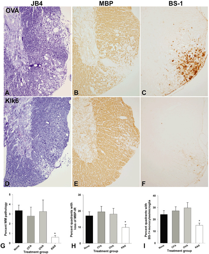
Decreased white matter pathology in the spinal cord of mice immunized with KLK6 in comparison to controls at 40 days post‐TMEV infection. A,D. Glycol methacrylate plastic‐embedded sections were stained with modified eriochrome/cresyl violet stain and the area of white matter pathology determined and expressed as a percent of total white matter (G). Parallel 1‐mm segments of spinal cord from the same mice were embedded in paraffin and 5‐µm sections stained for MBP (B,E) or for monocytes and activated microglia using a biotinylated BS‐1 antibody (C,F). The spinal cord of mice immunized with Klk6 5 weeks prior to TMEV infection showed significant reductions in the percent of white matter pathology (G, ANOVA, P = 0.009; *SNK P < 0.05) and in the percent of spinal cord quadrants exhibiting a loss of MBP staining (H, ANOVA on ranks, P = 0.03; *Dunn's P < 0.5) compared with immunization controls. Mice immunized with Klk6 prior to TMEV infection also showed fewer spinal cord quadrants associated with BS‐1‐positive monocytes and activated microglia (I, ANOVA, P = 0.003; *SNK, P < 0.05). See also Table 2.