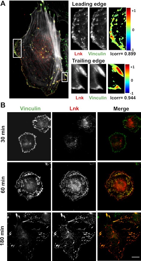
Figure 1.
Lnk colocalizes with adhesion sites in EC. A) HAECs were plated on glass coverslips, and confluent monolayers were wounded. After 2 h, the cells were fixed and immunostained for Lnk (red) and vinculin (green). Deconvolution of microscopic images was performed with Huygens Essential software. Colocalization colormap and correlation index (Icorr) were obtained as described in Materials and Methods, using ImageJ software. B) ECs were allowed to adhere to coated glass coverslips for various time periods (30, 60, and 180 min). The cells were fixed and immunostained for Lnk (red) and vinculin (green) and visualized as superimposition of images (merge). Scale bar = 10 μm.

